
Subject-specific vagus (SSV) scaffolds
This resource provides:
- A background of SPARC Phase 2 REVA initiative and associated datasets
- An overview of the workflows for generating subject-specific vagus (SSV) scaffolds from digitized vagus nerve segments
- Information on the coordinate systems and anatomical information contained within an SSV scaffold
- Tutorials for visualizing and analyzing SSV scaffolds
- Guidance on mapping SSV scaffolds into whole-body configurations
- Examples of using SSV scaffolds to investigate population-level nerve anatomy and nerve-organ relationships
- Links to published datasets, software and documentation
What can I do with an SSV scaffold?
SSV scaffolds provide a standardized three-dimensional representation of individual human vagus nerves while preserving subject-specific anatomical variation. They provide a common coordinate framework for integrating and comparing anatomical and multimodal data across subjects.
An SSV scaffold can be used to:
- Visualize anatomical relationships between the nerve trunk, branches and anatomical landmarks
- Characterize branch anatomy, including branch origins and initial directions
- Integrate multimodal data, including fascicular network representations and histological images
- Compare anatomy across samples using common vagus material coordinates
- Export anatomical representations for visualization, presentations, publications or computational analysis
- Generate anatomical flatmaps for organizing and exploring multimodal datasets
- Map into an in-body configuration to investigate nerve-organ relationships
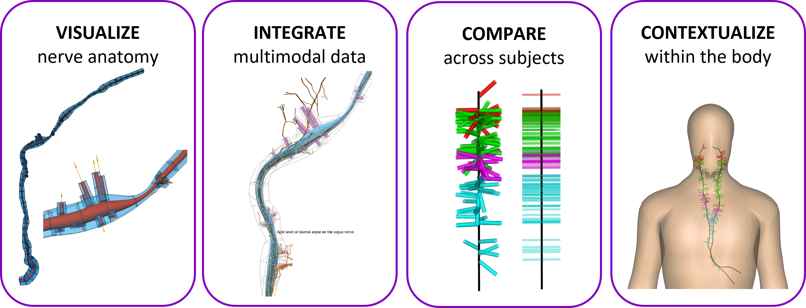
Figure 1. Main capabilities of SSV scaffolds. Together, these capabilities provide a common coordinate framework for studying human vagal anatomy.
SPARC Phase 2 REVA
The objective of SPARC Phase 2 REVA initiative is to characterize inter-subject variability in the human vagus nerve and provide insights into the detailed fascicular and microscopic structure and function of the nerve. The two REVA awardee teams - the Feinstein Institute for Medical Research & Temple University, and Case Western Reserve University & Duke University - have been producing high-resolution multimodal imaging and segmentation datasets of vagus nerves from a large cohort of human subjects.
For the purposes of this document, we focus on data from 29 cadavers that have been published by the Feinstein Institute as a collection of datasets (Human vagus nerve anatomical reconstruction using microCT, immunohistochemistry, and ultrasound).
The SPARC Data & Resource Center (DRC) works closely with the REVA teams to provide dataset curation and knowledge management, dataset storage, anatomical and functional mapping, simulation services, and access via the SPARC Portal.
Overview of the SSV scaffold generation workflow
An SSV scaffold is generated from digitized nerve segments from an ex-vivo specimen with the following workflows, which are implemented using the open-source Scaffold Mapping Tools:
- Data preparation
- Segmentation stitching
- SSV scaffold creation
The subject-specific vagus scaffolds are publicly available in Scaffold map - Human Vagus Nerve. The corresponding workflows are also available for each sample in the dataset's primary folder, with the output files from each workflow step in the derivative folder.
Data preparation
The raw data used to build the vagus nerve scaffolds are derived from ex-vivo specimens that REVA teams cut into segments for micro-CT imaging, followed by digitization and annotation of anatomical features and orientations, as shown in Figure 2. These annotated segmentation files can typically be found in REVA datasets in either XML or CSV format.
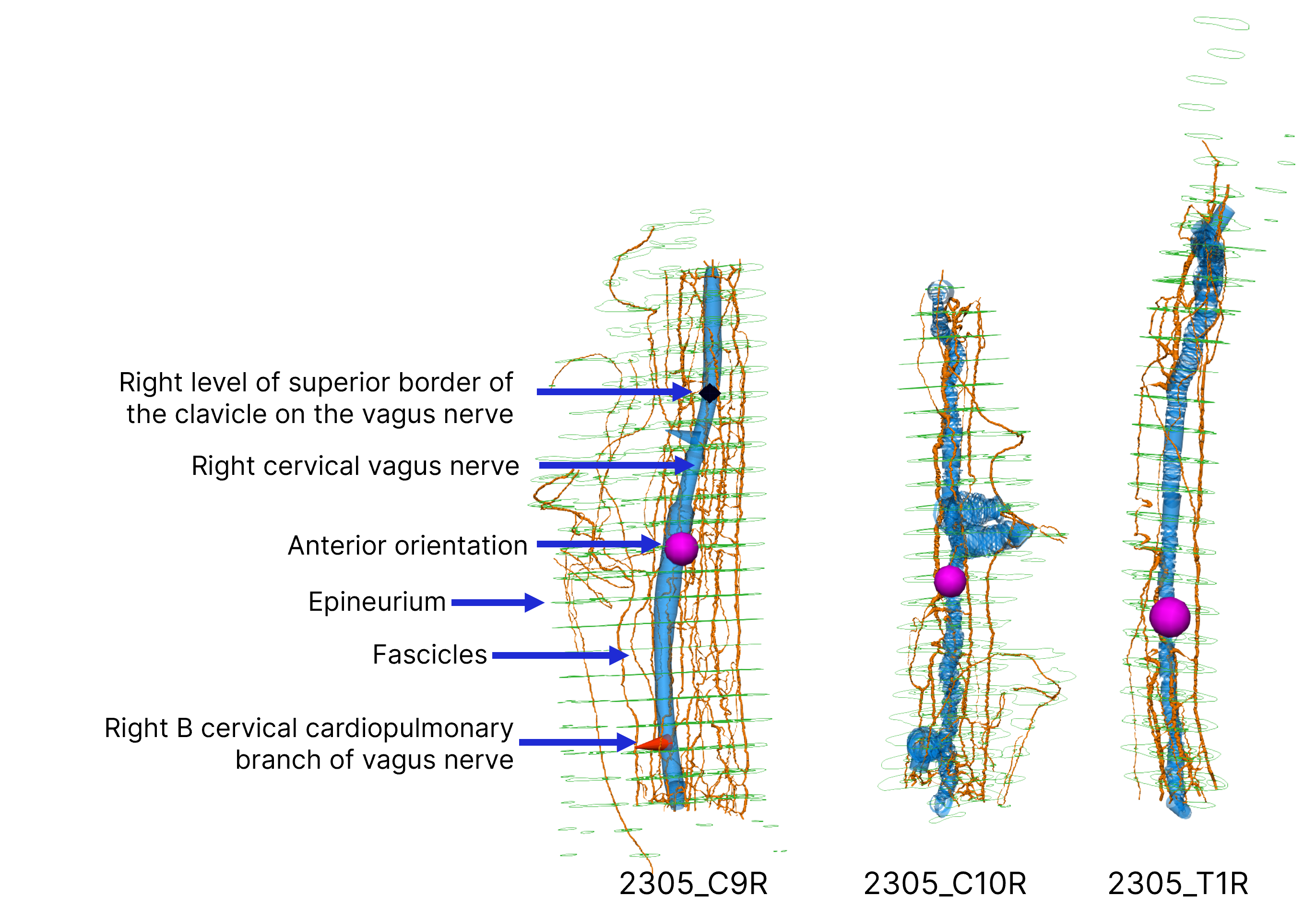
Figure 2. Annotated digitized segments from subject 2305 (DOI: 10.26275/imas-52lf). The magenta sphere represents the anterior orientation of each segment.
For each sample, its digitized segments can be retrieved directly from the SPARC Portal using the Retrieve Portal Data plugin and the DOI of the sample dataset. The retrieved segmentation files are then converted to EXF format, which allows them to be ingested by the Scaffold Mapping Tools. The data preparation workflow is shown in Figure 3.
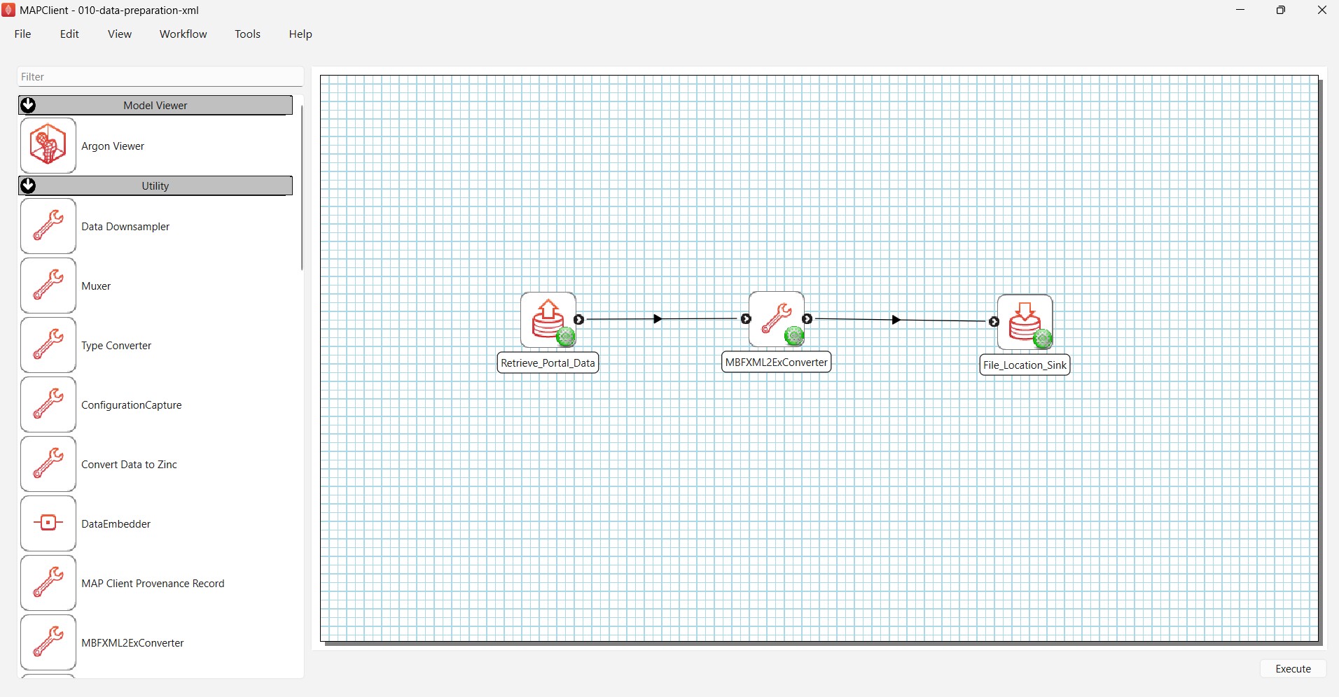
Figure 3. Data preparation workflow for retrieving data files from the SPARC Portal and converting them into a format suitable for use with the Scaffold Mapping Tools.
Segmentation stitching
The digitized segments from each sample nerve are stitched together to form a continuous nerve in a non-body configuration using a semi-automated process. This step is performed with the Segmentation Stitcher plugin from the Scaffold Mapping Tools as shown in Figure 4.
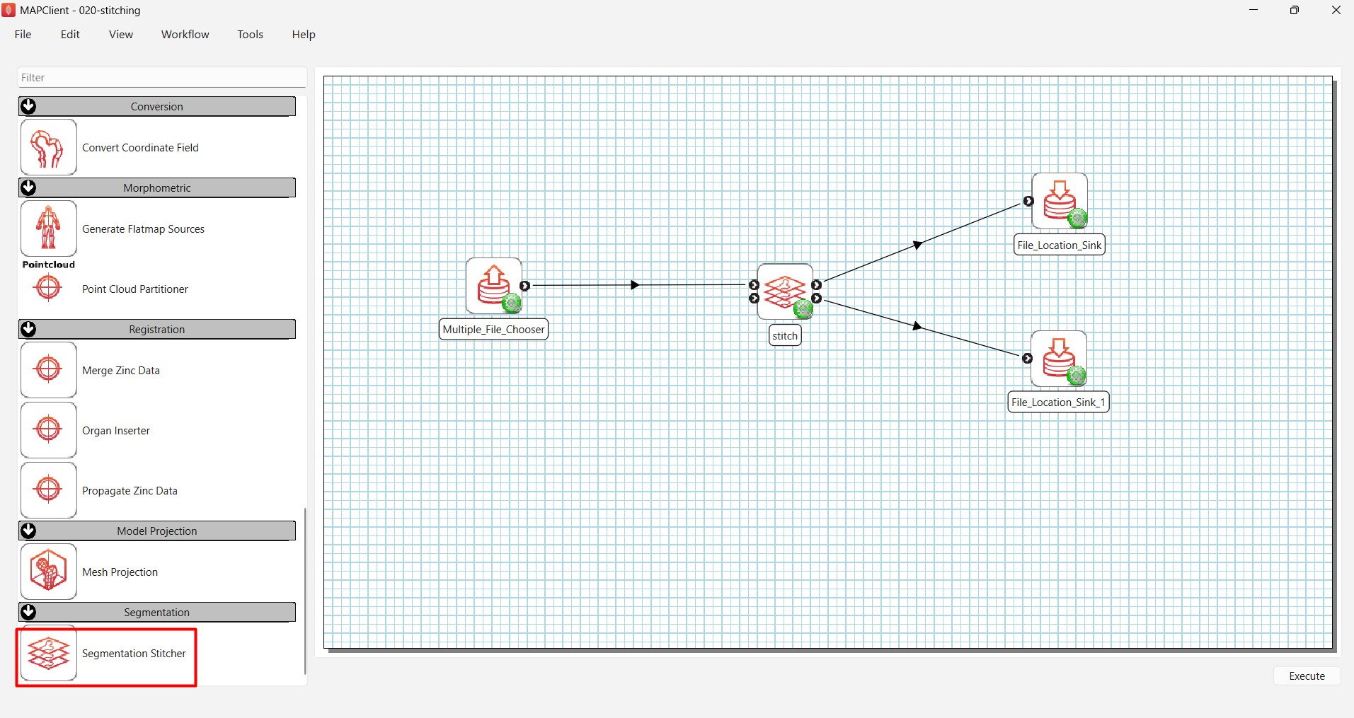
Figure 4. Workflow for stitching digitized nerve segments using the Segmentation Stitcher (red box).
The Segmentation Stitcher reads all segment files for a sample and provides an interactive environment for positioning the segments in the correct anatomical order, as shown in Figure 5. The fascicles (orange) and nerve trunk (blue) on the end face of each segment are then joined to their corresponding fiber on the start face of the next segment. Connections can be made using the built-in shape-matching optimization algorithm or manually using fascicle correspondence information provided by the REVA teams.
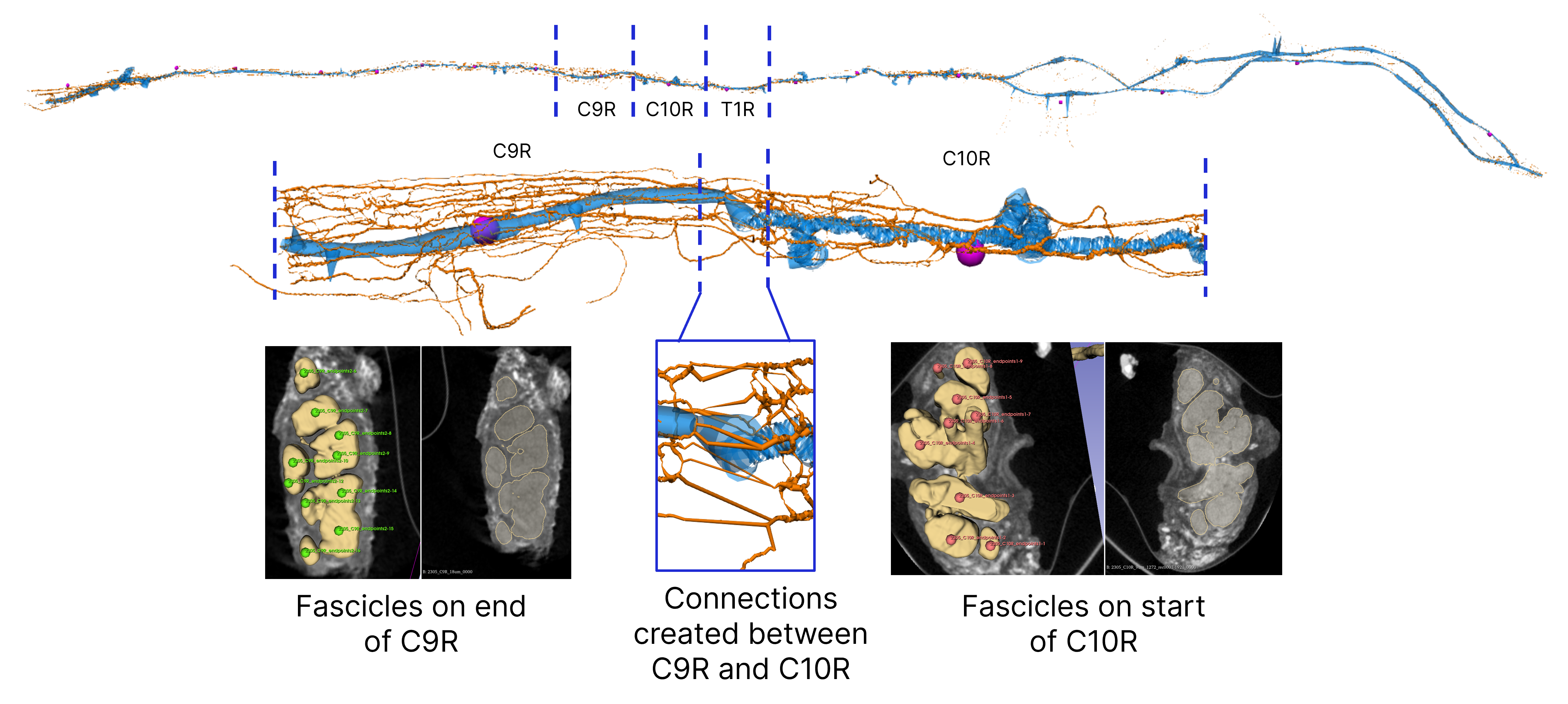
Figure 5. Stitching of cut segments using the Segmentation Stitcher.
The result of this step is a continuous representation of the vagus nerve that preserves the anatomical features and branching information contained in the original digitized segments.
SSV Scaffold creation
The Scaffold creator is then used to fit a standardized nerve trunk to the path, radius, anatomical landmarks and orientation of the stitched data, as shown in Figure 6.
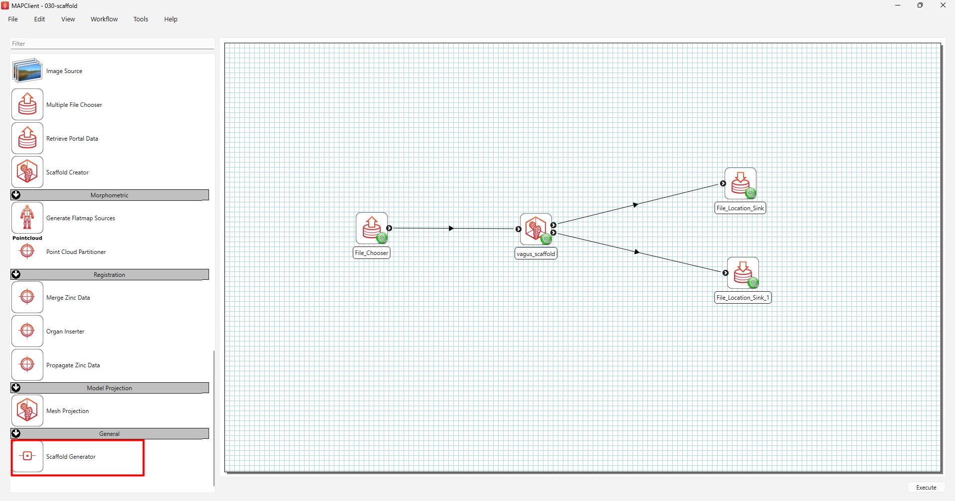
Figure 6. Workflow for generating a SSV scaffold by fitting a standardized nerve trunk to the stitched data using the Scaffold Creator (red box).
The resulting SSV scaffold is a volumetric finite element model of the individual nerve. Branching information contained in the stitched data is captured during the fitting process. Branch starting locations and initial directions are embedded within the trunk volume, as shown in Figure 7. Anatomical feature annotations from the stitched data are also transferred to the scaffold.
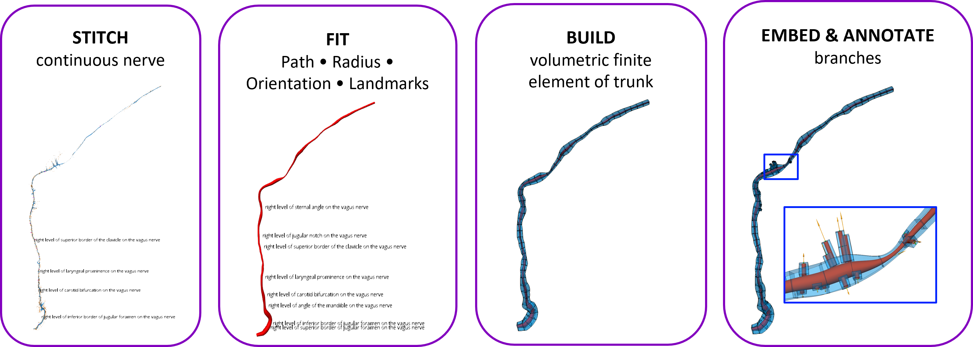
Figure 7. An SSV scaffold is generated by fitting a trunk to the nerve path, radius, orientation and anatomical landmarks of the stitched data, followed by building a volumetric representation of the nerve trunk. Starting locations and initial directions of branches are then embedded in the trunk volume in an SSV scaffold.
Each SSV scaffold is defined with three coordinate fields: coordinates, straight coordinates and vagus coordinates, as shown in Figure 8.
coordinatesdefine the scaffold's geometry and are typically derived from stitched data.straight coordinatesprovide the corresponding geometric coordinates as if the same nerve trunk were laid out smoothly along a straight axis without twists. This coordinate system facilitates comparison of the gross anatomy of different nerve samples.vagus coordinatesare material coordinates that provide a normalized measure down the vagus nerve, ranging from 0.0 near the brainstem to 1.0 at the end of the scaffold. Anatomical level markers are calibrated at fixed locations along the nerve centroid.
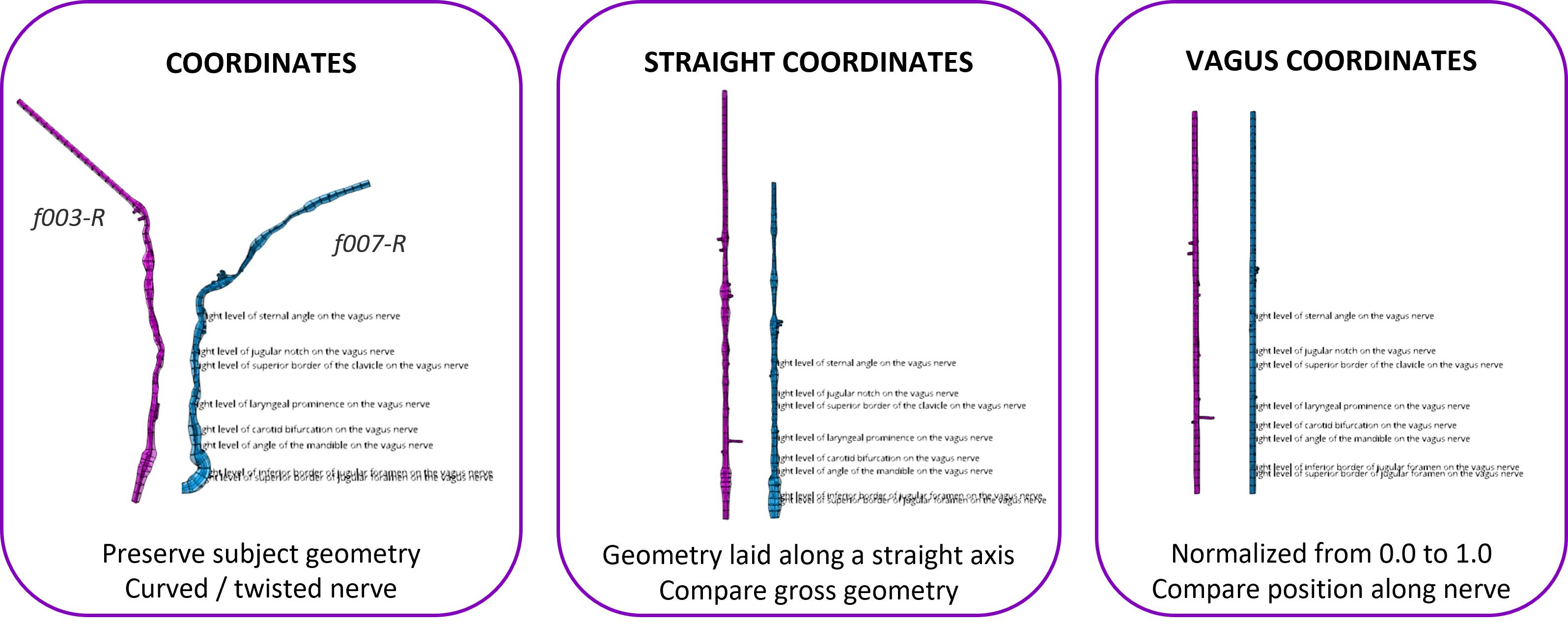
Figure 8. The three coordinate systems defined by an SSV scaffold.
The common vagus coordinates are particularly important for population-level analysis. Although nerve trunk lengths and branching patterns vary substantially between individuals, the normalized coordinate system allows anatomical features from different subjects to be mapped onto a common reference space.
Further technical information about the vagus nerve scaffold is available in the Vagus Scaffold Documentation.
Using subject-specific vagus scaffolds
Population-level analysis
The vagus coordinates allow anatomical features from multiple SSV scaffolds to be compared directly. For example, branch origins and initial directions can be mapped from individual subjects into the common vagus coordinate system to investigate inter-subject variation.
Figure 9(i) shows branch origins and initial directions of SSV scaffolds generated from 20 subjects in the Feinstein collection, where the branches are mapped to the vagus coordinates for qualitative comparison. A simplified representation of branch origin points, shown in Figure 9(ii), provides a direct way to visualize the clustering and overlap among different branch groups.
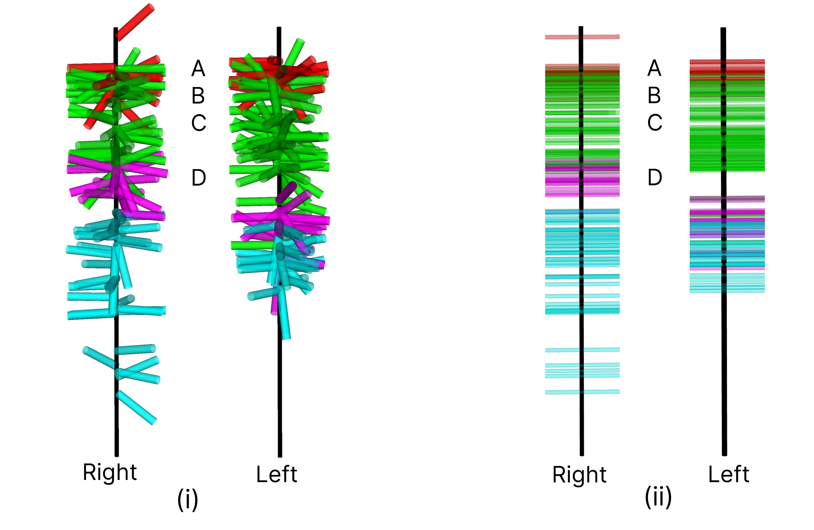
Figure 9. SSV scaffolds from 20 subjects in the Feinstein collection mapped to vagus coordinates. Superior laryngeal nerve branches are represented in red, cardiovascular branch of cervical vagus nerve in green, recurrent laryngeal nerve in magenta and esophageal branch of esophageal plexus of vagal nerve in cyan. The level of the inferior border of the jugular foramen, the level of the carotid bifurcation, the level of the laryngeal prominence and the level of the superior border of the clavicle are represented by levels A-D, respectively.
The steps for creating such visualizations are described in Scaffold Mapping Tools: Using Subject-Specific Vagus (SSV) Scaffolds For Population-Level Analysis. The SPARC-SSV tutorials also provide examples of how to work with SSV scaffolds and extract key information, such as structure maps, branch starting locations and initial directions, for downstream analysis.
Visualization and export
Each SSV scaffold and its associated data can be visualized on the SPARC Portal through the dataset Gallery. The ScaffoldVuer allows users to visualize the scaffold together with the stitched data, as shown in Figure 10. The Regions panel in the bottom-left corner can be used to toggle the visibility of individual graphic objects and interrogate the relationship between the scaffold and the source data.
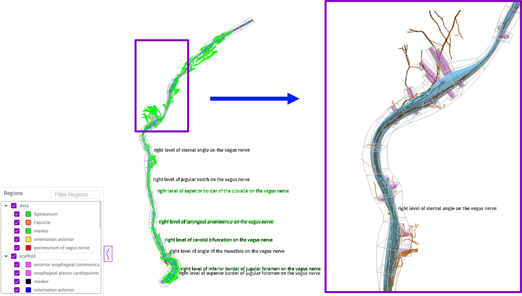
Figure 10. Gallery view of an SSV scaffold derived from f007-right (DOI: 10.26275/dp8r-bb7w), with a zoom-in view of the fascicles and vagal branches.
SSV visualizations are generated using Argon Viewer and exported in multiple format using the Argon Scene Exporter plugins, as shown in the workflow in Figure 11.
The scaffold can be exported in:
- WebGL format, for interactive visualization through the SPARC Portal Gallery
- PNG image format for presentation or publication
- STL, VTK, and MBFXML formats for reuse in other applications
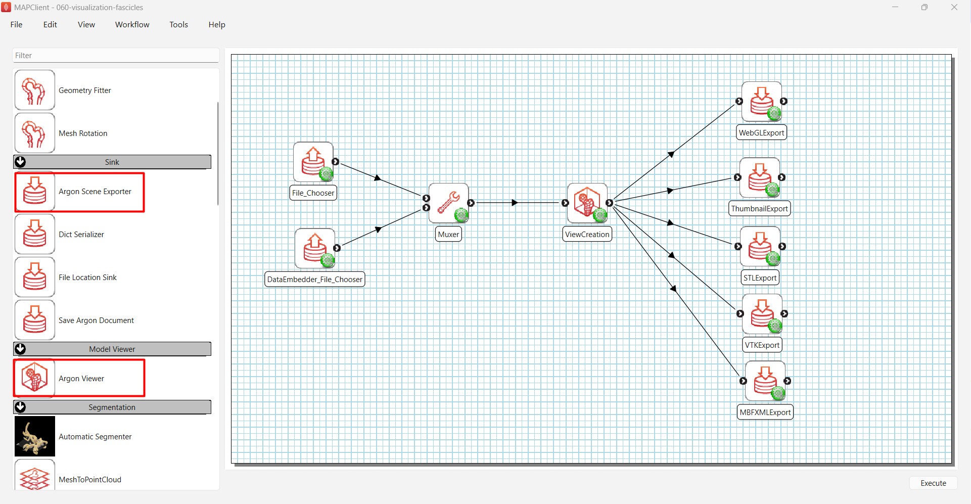
Figure 11. Workflow for generating and exporting visualizations of SSV scaffolds and associated stitched data.
Flatmap generation
A SSV scaffold can be flattened into an anatomical map using mapmaker. The resulting flatmap is an SVG drawing together with its associated metadata, as shown in Figure 12.
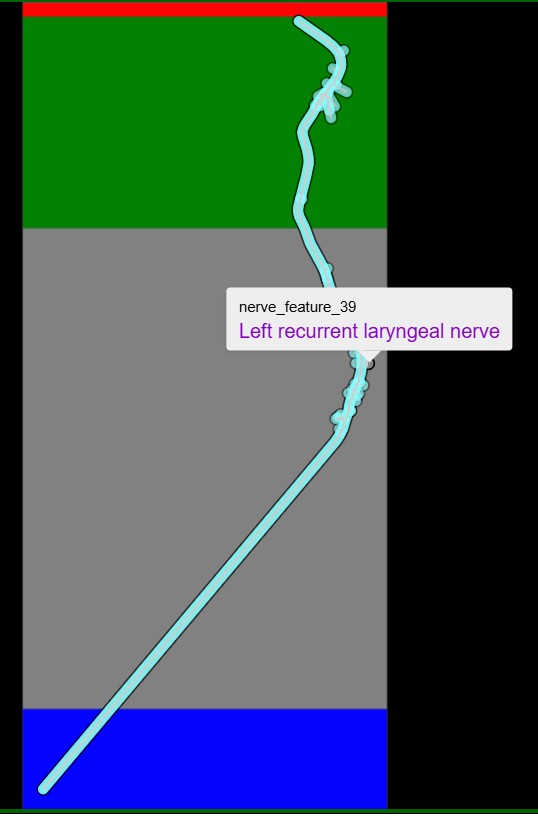
Figure 12. Flatmap of the left SSV generated from f008 (DOI: 10.26275/gpmp-nogs). The vagus nerve trunk, branches and level markers are annotated in the flatmap. The brainstem region is represented by the red band, the cervical region by the green band, the thoracic region by the grey band and the lumbar region by the blue band.
Generated flatmaps are ingested into the Human Vagus Nerve Explorer. Each flatmap provides a spatial framework for referencing histological images and other multimodal data collected for the corresponding sample.
In-body configurations
SSV scaffolds can be remapped into an in-body configuration using subject-specific digitized data or inserted into a reference whole-body scaffold containing an oriented nerve trunk. The procedure for inserting SSV scaffolds into a whole-body scaffold is described in detail in Scaffold Mapping Tools: Inserting Subject-Specific Vagus (SSV) Scaffolds In Whole-Body Scaffold.
Mapping SSV scaffolds into a common body configuration enables population-level analysis of nerve anatomy in an anatomical context. Figure 13 shows 40 SSV scaffolds from the Feinstein collection mapped onto a reference whole-body scaffold. Branch origins and initial directions can be visualized across subjects, while individual subjects can be highlighted with gold spheres for comparison.
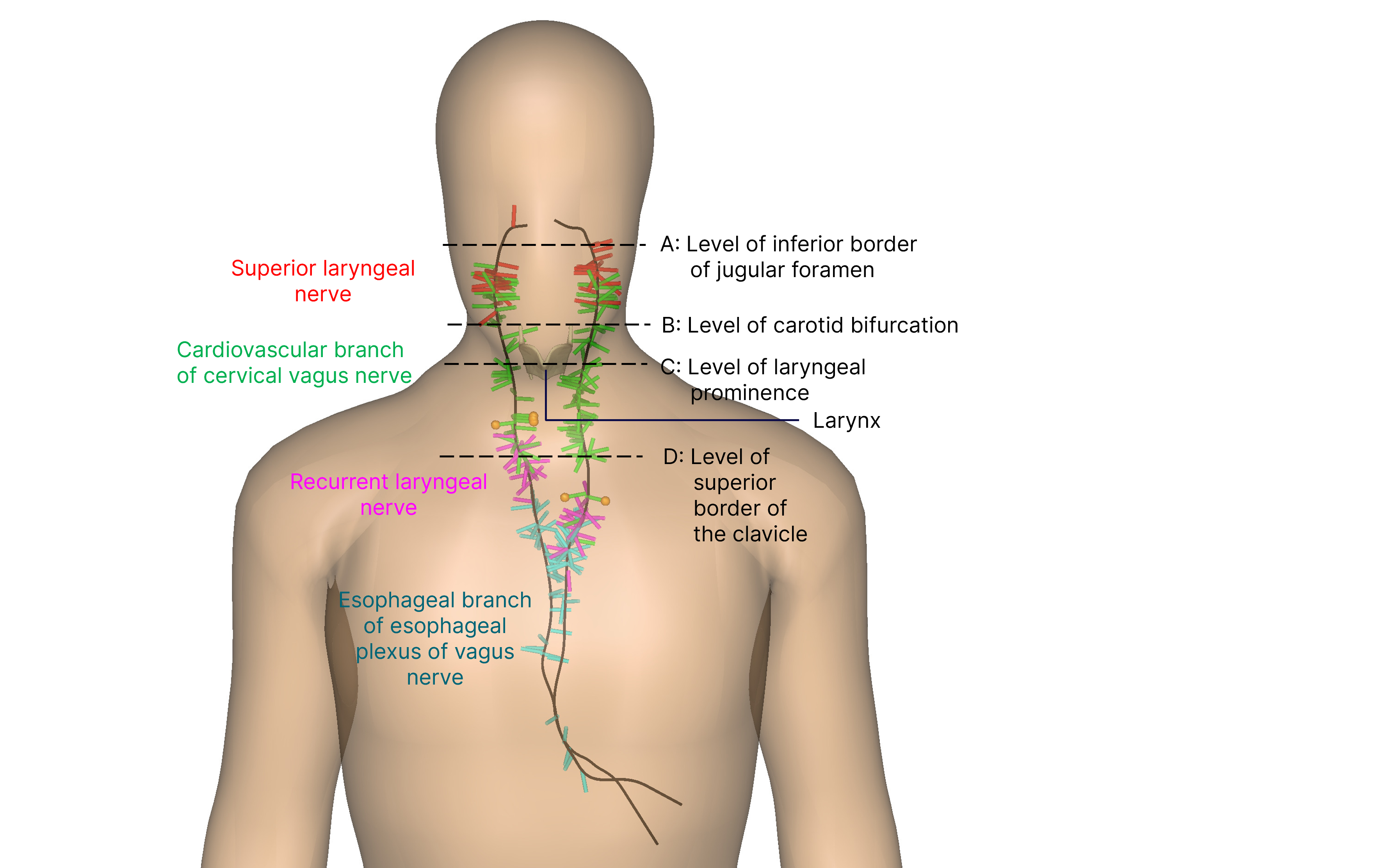
Figure 13. Insertion of 40 SSV scaffolds derived from the Feinstein collection into a reference whole-body scaffold.
The whole-body scaffold framework can also incorporate existing organ scaffolds, such as the larynx, as shown in Figure 13, allowing the spatial relationships between vagal branches and their target organs to be examined.
By mapping specific vagal branch groups across multiple subject-specific vagus scaffolds and visualizing them together with target organs scaffolds, users can investigate how branches are distributed along the nerve trunk and how they extend and interact spatially with their target organs. This provides a framework for studying nerve-organ relationships at the population level and has potential applications in understanding vagal anatomy, investigating functional innervation patterns and informing the design and placement of stimulation devices.
Datasets and Resources
| Resources | Description |
|---|---|
| SPARC Phase 2 REVA | Background and information about the REVA initiative |
| Human vagus nerve anatomical reconstruction using microCT, immunohistochemistry, and ultrasound | A published collection of 29 datasets from the Feinstein group, source data for SSV scaffolds |
| Scaffold map - Human Vagus Nerve | Published SSV scaffolds, workflows and derivative files |
| SPARC-SSV tutorials | Collection of tutorials in the form of iPython Jupyter notebooks for working with SSV scaffolds |
| Scaffold Mapping Tools: Using subject-specific vagus (SSV) scaffolds for population-level analysis | Guide for visualizing SSV scaffolds in vagus material coordinates for population studies |
| Scaffold Mapping Tools: Inserting Subject-Specific Vagus (SSV) Scaffolds In Whole-Body Scaffold | Guide for insertion of SSV scaffolds in reference whole-body scaffold |
| Vagus Scaffold Documentation | Technical reference for the vagus scaffold |
| Human Vagus Nerve Explorer | Dashboard for exploration of REVA data |
| Scaffold Mapping Tools | Open-source software tool for generating SSV scaffolds |
Summary
Subject-specific vagus scaffolds provide a common anatomical framework for representing and comparing the human vagus nerve while preserving subject-specific variation. Starting with digitized excised nerve segments, the workflow produces a volumetric scaffold that contains the nerve trunk, anatomical landmarks and branching information in a standardized coordinate system.
Once generated, these scaffolds can be used for population-level anatomical analysis, multimodal integration, visualization, flatmap generation, and mapping into whole-body configurations. Integration with organ scaffolds in the whole-body framework enables investigation of the spatial relationships between vagal branches and their target organs.
These capabilities provide a reproducible framework for moving from high-resolution subject-specific vagus nerve data to population-level anatomical analysis and exploration of nerve-organ interactions.
Updated about 2 months ago